
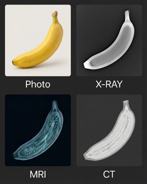
At first glance, a banana seems like an unlikely teaching tool for medical imaging—but that’s exactly what makes it effective. The image above shows a banana captured using standard photography alongside X-ray, CT, and MRI, offering a simple, intuitive comparison of how each modality “sees” the same object.
Plain Photo: What the Eye Sees
The photograph represents our baseline: surface color, shape, and texture. While useful for orientation, it tells us nothing about internal structure—precisely where medical imaging adds value.
X-ray: Density in Its Simplest Form
In the X-ray image, the banana appears mostly uniform with subtle variations. X-ray imaging is driven by differences in attenuation, so soft tissues with similar densities tend to blend together. This mirrors clinical reality: X-ray excels at detecting high-contrast structures (like bone or metal), but offers limited soft-tissue detail.
CT: Cross-Sectional Density Mapping
The CT image adds clarity by reconstructing attenuation data into cross-sectional detail. Here, the banana’s peel and inner pulp become distinguishable due to small density differences. This highlights CT’s strength in resolving subtle contrast while maintaining excellent spatial resolution—one reason it’s so widely used for rapid, whole-body assessment.
MRI: Signal, Not Density
The MRI view looks dramatically different. Instead of density, MRI reflects differences in tissue properties such as proton density and relaxation times. The internal fibrous structure of the banana becomes visible, illustrating why MRI is so powerful for soft-tissue characterization—and why its images often look unfamiliar to those trained primarily on X-ray or CT.
Why This Matters for Imaging Professionals
Using a familiar object removes anatomy from the equation and puts the focus squarely on physics and modality strengths. For continuing education, examples like this reinforce a core principle of medical imaging: each modality answers different clinical questions. Mastery comes from understanding not just how to acquire images, but how and why they look the way they do.
Sometimes, the best way to sharpen our diagnostic perspective is to look at something completely ordinary—through an extraordinary lens. 🍌